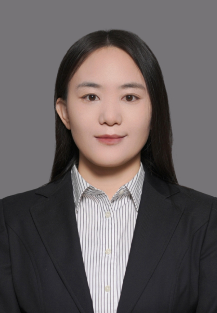
Kunrong Mei——Associate Professor

Nationality:
Chinese
Phone:
15022575215
Email:
kmei@tju.edu.cn
Office:
Room 417-2, Building 24, Tianjin University
School:
School of Pharmaceutical Science and Technology
ResearcherID:
Group weblink
Education Experience
2005-2009 Bachelor of Engineering Bioinformatics Huazhong University of Science and Technology
2009-2014 Doctor of Philosophy Structural Biology Tsinghua University
Professional Experience
2014-2019 Postdoc University of Pennsylvania
2019- Associate professor Tianjin Univesity
Research Area

    We’re interested in solving important and interesting scientific problems with structural biology and protein engineering techniques. Currently, our research has been focused on three directions: 1) Molecular mechanism underlying the regulation of vesicle trafficking. 2) Structure based semi-rational engineering of enzymes. 3)Orthogonal system in yeast cells.

1)  Molecular mechanism underlying the regulation of vesicle trafficking.

   Vesicle trafficking is a fundamental cellular process by which membrane-encapsulated vesicles transport materials between different cellular compartments and between a cell and its environment. Malfunction of vesicle trafficking will cause a broad spectrum of severe diseases, such as cancer, diabetes, immune deficiency and neuropathy. Vesicle trafficking occurs in four steps including vesicle biogenesis, transport, docking and fusion with the target membrane, each of which is mediated by a specific concert of protein families. Membrane fusion is mediated by the SNAREs and docking is manly mediated by the MTCs (Multi-subunit tethering complexes). MTCs not only mediate vesicle docking via tethering the trafficking vesicles to the target membrane, but also interact with the SNAREs to promote membrane fusion between them (Figure 1). However, the molecular mechanisms underlying these cellular processes are largely unknown.

Figure 1. MTCs and SNAREs in different stages of vesicle trafficking. MTCs are shown in light blue boxes, and SNAREs in white boxes.


With multidisciplinary strategies including structural biology, biochemistry, cell biology and yeast genetics, we have uncovered the molecular mechanism of exocyst-assembly (Figure 2, ref 1). We continue to work on the functional mechanism of other MTCs, as well as the interaction between MTCs and SNAREs.

Figure 2. The hierarchical assembly of the exocyst complex. The eight exocyst subunits share similar structural folds, with an N-terminal CorEx motif (the blue coiled-coil) followed by a long helical rod to the C-terminus (top left). Sec3 and Sec5, Sec6 and Sec8, Sec10 and Sec15, Ex70 and Exo84 form heterodimers through the pairing of their CorEx motifs (down left). Then Sec3, Sec5, Sec6 and Sec8 form the Subcomplex I via four helix bundle formation of their CorEx motifs; Sec10, Sec15, Exo70 and Exo84 form the Subcomplex II in the same manner (down right). Finally, Subcomplex I and Subcomplex II clap into each other to form the fully assembled exocyst complex (top right).


2) Structure based semi-rational engineering of enzymes.

     The biosynthesis of many important nature products, e.g. ginsenoside, is mediated by plant enzymes. Since the production of these nature products from native plants are often quite low, it is necessary to improve their biosynthesis via bio-techniques. Structure based semi-rational design is a good way to achieve it. Key residues of the enzymes involved in catalysis of substrates could be analyzed based on the structure or predicted structure and known knowledge. And improved enzymes may be obtained via a series of mutations of these residues. In this field, our research includes both structure determination and structure-based engineering of interested enzymes (Figure 3, ref 2).


Figure 3. Semi-rational design of Pq3-O-UGT2. A) The structure model of Pq3-O-UGT2 is superimposed onto crystal structure of UGT74AC1. Mutant residues of engineered UGT74AC1 that highly improved its activities and corresponding residues of Pq3-O-UGT2 are indicated. B) Catalytic conformations of Pq3-O-UGT2 (WT)-UDPG-Rh2 (top) and Pq3-O-UGT2 (S49R/I50M/H85Y, Mutant)-UDPG-Rh2 (bottom) after MD simulations. The mutations improve the interaction between Rh2 and Pq3-O-UGT2.


3) Orthogonal system in yeast cells.

   Recently, a group has reported application of linear plasmids in budding yeast to obtain autonomous hypermutation of interested proteins, including nanobodies and enzymes (Figure 4, ref 3). We are very interested in this powerful and cost-effective system. Thus, right now, we are trying to set up this system in our lab and using it to obtain desired mutations of proteins we are interested in.


Figure 4. An orthogonal replication system based on the p1/2 replication system. The replication of p1/2 linear plasmids is mediated by TP-DNAP1 and TP-DNAP2, which is independent of host DNA polymerases. Thus, mutations could be introduced to p1/2 via TP-DNAP1 during proliferation of the host cells. 

Reference

1. Mei K, Li Y, Wang S, Shao G, Wang J, Ding Y, Luo G, Yue P, Liu JJ, Wang X, Dong MQ, Wang HW, Guo W. Cryo-EM structure of the exocyst complex. Nat Struct Mol Biol. 2018 Feb;25(2):139-146.

2. Yao L, Zhang H, Liu Y, Ji Q, Xie J, Zhang R, Huang L, Mei K, Wang J, Gao W. Engineering of triterpene metabolism and overexpression of the lignin biosynthesis gene PAL promotes ginsenoside Rg3 accumulation in ginseng plant chassis. J Integr Plant Biol. 2022 Jun 22. doi: 10.1111/jipb.13315. Epub ahead of print.

3. Ravikumar A, Arrieta A, Liu CC. An orthogonal DNA replication system in yeast. Nat Chem Biol. 2014 Mar;10(3):175-7. 

Honors and Awards
Patents
Highlighted Publications
1. Liu, Y., Li, M., You, X., Ji, Q., and Mei, K. Advances in understanding the structure and function of the exocyst complex. Sci. Sin.-Vitae. 2022, 52, 95–106
2. Yao L, Zhang H, Liu Y, Ji Q, Xie J, Zhang R, Huang L, Mei K, Wang J, Gao W. Engineering of triterpene metabolism and overexpression of the lignin biosynthesis gene PAL promotes ginsenoside Rg3 accumulation in ginseng plant chassis. J Integr Plant Biol. 2022 Jun 22. doi: 10.1111/jipb.13315. Epub ahead of print. PMID: 35731022.
3. Huang H, Ouyang Q, Mei K, Liu T, Sun Q, Liu W, Liu R. Acetylation of SCFD1 regulates SNARE complex formation and autophagosome-lysosome fusion. Autophagy. 2022 Apr 24:1-15.
4. Mei K, Liu DA, Guo W. Determine the Function of the Exocyst in Vesicle Tethering by Ectopic Targeting. Methods Mol Biol. 2022;2473:65-77.
5. Huang H, Ouyang Q, Zhu M, Yu H, Mei K, Liu R. mTOR-mediated phosphorylation of VAMP8 and SCFD1 regulates autophagosome maturation. Nat Commun. 2021 Nov 16;12(1):6622.
6. Liu S, Fu Y, Mei K, Jiang Y, Sun X, Wang Y, Ren F, Jiang C, Meng L, Lu S, Qin Z, Dong C, Wang X, Chang Z, Yang S. A shedding soluble form of interleukin-17 receptor D exacerbates collagen-induced arthritis through facilitating TNF-α-dependent receptor clustering. Cell Mol Immunol. 2021 Aug;18(8):1883-1895.
7. Mao, L., Zhan, Y.-Y., Wu, B., Yu, Q., Xu, L., Hong, X., Zhong, L., Mi, P., Xiao, L., Wang, X., Cao, H., Zhang, W., Chen, B., Xiang, J., Mei, K., Radhakrishnan, R., Guo, W., and Hu, T. ULK1 phosphorylates Exo70 to suppress breast cancer metastasis. Nat Commun. 2020, 11, 117
8. K. Mei and W. Guo. Exocytosis: A New Exocyst Movie. Current Biology, 2019, 29(1), R30-R32.
9. K. Mei, P. Yue,and W. Guo. Analysis of the Role of Sec3 in SNARE Assembly and Membrane Fusion. SNAREs, 175-189. (Springer New York, 2019)
10. K. Mei and W. Guo. The exocyst complex. Current Biology, 2018, 28(17), R922-R925.
11. K. Mei*, Y. Li*, S. Wang, G. Shao, J. Wang, Y. Ding, G. Luo, P. Yue, J.-J. Liu, X. Wang, M.-Q. Dong, H.-W. Wang, and W. Guo. Cryo-EM Structure of the Exocyst Complex, Nature structural & molecular biology, 2018, 25(2):139-146.
12. P. Yue*, Y. Zhang*, K. Mei, S. Wang, J. Lesigang, Y. Zhu, G. Dong, and W. Guo. Sec3 Promotes the Initial Binary T-Snare Complex Assembly and Membrane Fusion, Nature Communication, 2017, 8:14236.
13. S. Tang, Y. Si, Z. Wang, K. Mei, X. Chen, J. Cheng, J. Zheng, and L. Liu. An Efficient One‐Pot Four‐Segment Condensation Method for Protein Chemical Synthesis, Angewandte Chemie International Edition, 2015, 54(19): 5713-5717.
14. S. Yang*, Y. Wang*, K. Mei, S. Zhang, X. Sun, F. Ren, S. Liu, Z. Yang, X. Wang, Z. Qin, and Z. Chang. Tumor necrosis factor receptor 2 (TNFR2)· interleukin-17 receptor D (IL-17RD) heteromerization reveals a novel mechanism for NF-κB activation, Journal of Biological Chemistry, 2015, 290(2): 861-871.
15. K. Mei*, Z. Jin*, F. Ren, Y. Wang, Z. Chang, and X. Wang. Structural Basis for the Recognition of RNA Polymerase II C-Terminal Domain by CREPT and p15RS, Science China Life Sciences, 2014, 57(1): 97-106.
16. D. Wang, S. Zhang, L. Li, X. Liu, K. Mei, and X. Wang. Structural insights into the assembly and activation of IL-1 [beta] with its receptors, Nature immunology, 2010,11(10): 905-911.